Introduction to 3D Sonograms
Expecting a baby is one of the most exciting times in a parent’s life. With today’s advanced imaging technology, parents can get an incredibly detailed look at their growing baby with 3D sonograms. Unlike traditional ultrasound images, 3D sonograms provide a more lifelike, three-dimensional view of the baby. This gives parents a clearer picture of the baby’s face, hands, and other tiny features, allowing for a special bonding experience even before birth.
How 3D Sonograms Differ from 2D Ultrasounds
A 2D ultrasound captures flat, black-and-white images of the baby. While it can show the baby’s position and general anatomy, it lacks depth and detail. In contrast, a 3D sonogram uses advanced technology to combine multiple 2D images. This creates a three-dimensional view that highlights the baby’s contours and features in much greater detail. Parents can now see expressions, tiny hands, and even the shape of their baby’s nose, making the experience more personal and memorable.
Benefits of 3D Sonograms
3D sonograms offer several advantages over traditional ultrasounds. First, they provide a clear and realistic image that can make parents feel more connected to their baby. Seeing detailed images can also reduce parental anxiety by confirming normal development. Additionally, doctors can use 3D sonograms to detect certain facial and skeletal abnormalities early, allowing for timely intervention. These benefits make 3D imaging a valuable tool for both medical and personal purposes.
The Science Behind 3D Imaging
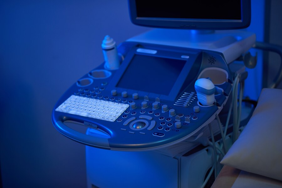
3D sonograms use high-frequency sound waves, like traditional ultrasounds. The difference lies in the software that processes multiple angles of 2D images and renders them into a 3D model. The sonographer applies a special probe that moves along the mother’s abdomen, capturing different views of the baby. The software then combines these views into a single 3D image. This process is safe, non-invasive, and provides clear, detailed images of the baby.
When is the Best Time for a 3D Sonogram?
Most healthcare providers recommend scheduling a 3D sonogram between 26 to 32 weeks of pregnancy. At this stage, the baby has developed recognizable features, but there’s still enough amniotic fluid to capture clear images. In earlier weeks, the baby’s features may not be as defined. Later, the baby may be too large and in a position that obstructs clear imaging, making this window optimal for capturing detailed sonogram images.
How to Prepare for Your 3D Sonogram Appointment
Preparing for a 3D sonogram appointment is straightforward but can improve image quality. Staying hydrated for a few days before the appointment can help by increasing the clarity of the amniotic fluid around the baby. Some sonographers also suggest eating a light snack or drinking something cold right before the session to make the baby more active. This can encourage the baby to move into a position that allows for better imaging.
What to Expect During the Appointment
A 3D sonogram appointment is similar to a regular ultrasound appointment. The sonographer will apply gel to the mother’s abdomen, which helps the probe pick up sound waves more effectively. The probe moves over the belly, capturing multiple images. These images are processed instantly, allowing parents to see the baby’s features in real-time. The appointment is comfortable, non-invasive, and usually lasts about 20-30 minutes.
Are 3D Sonograms Safe?
Yes, 3D sonograms are considered safe for both mother and baby. The technology used in 3D imaging is the same as that in traditional ultrasounds, which have been used for decades without harmful effects. The main difference lies in the processing software, not the sound waves themselves. Medical experts agree that, when performed by trained professionals, 3D sonograms are safe and pose no known risks.
Emotional Impact of 3D Sonograms on Parents
3d ultrasound have an undeniable emotional impact on parents, making the experience deeply personal. Seeing their baby’s face, fingers, and toes often evokes strong emotions. Many parents report feeling a stronger connection with their baby and describe the experience as an unforgettable bonding moment. The lifelike images also provide a tangible sense of the baby’s personality and appearance, which can deepen the emotional journey of pregnancy.
The Cost of 3D Sonograms and Insurance Coverage
The cost of a 3D sonogram can vary widely depending on the location and the imaging center. On average, parents can expect to pay between $100 to $300. Some insurance policies cover 3D imaging if it is medically necessary. However, if it is done solely for keepsake purposes, it is usually considered an out-of-pocket expense. Checking with the provider and insurance company before scheduling can clarify any coverage details.
Common Misconceptions About 3D Sonograms
There are some misconceptions surrounding 3D sonograms. One common myth is that they use more intense sound waves, which is not true. 3D sonograms use the same frequency and strength of sound waves as standard ultrasounds. Another misconception is that 3D imaging is only for keepsakes; in fact, it has significant medical applications. Doctors can use 3D sonograms to monitor certain conditions and detect developmental issues early.
Choosing a Reliable 3D Sonogram Provider
Choosing a reliable provider for a 3D sonogram is essential for a safe and rewarding experience. Look for certified technicians and facilities with modern equipment. Checking online reviews and asking for recommendations from your healthcare provider can also help. Ensuring that the sonogram is done by a licensed professional minimizes any risks and enhances the quality of the images, giving parents a clear view of their baby.
The Future of Sonogram Technology
Sonogram technology continues to evolve, with 4D imaging now available in some facilities. This new technology adds real-time movement, allowing parents to watch their baby’s motions live. As imaging software advances, the quality and accessibility of 3D and 4D sonograms will likely improve. In the future, these technologies may even allow for more detailed medical assessments and new ways for parents to experience their baby’s development.
Conclusion: The Unique Joy of 3D Sonograms
3D sonograms offer a unique way for parents to connect with their growing baby. The detailed images provide a memorable experience and can bring peace of mind about the baby’s health. By choosing a reputable provider and preparing properly, parents can enjoy the full benefits of this technology. As imaging technology advances, 3D sonograms will continue to play an important role in both prenatal care and the joyful experience of expecting a child.


